Showing posts with label 3D printing. Show all posts

Saturday, September 10, 2016

3D food printing technology : 3D printing with cow DNA.

Perfect Day Foods they’ve found a way to replicate cow DNA through 3D printing, fabricating a tasty alternative that is vegan, and both lactose- and gluten-free. Perfect Day Foods founded in 2014, isn’t just the whimsical concept of two hungry dreamers. They’ve worked to raise a total of $4 million in investment funds for the 3D printing of this bovine DNA and subsequent new dairy. This entrepreneurial team is planning to give us our ice cream back with a substitute that they feel confident we’ll enjoy much more than all the other ones we pass by in the grocery store today with a mental ‘ick.’ 

                      3D food printing with  cow DNA.
 
This apparently is not about the all too common labeling and trendy vegetarian marketing that still, ultimately, means trying to force something overpriced, frightening, and weird down our deprived little gullets. Perfect Day’s animal-free dairy milk is a product created by people as hungry as you and I are, clamoring for our cheese. And in fact, it’s so close to the real thing—even made with casein and whey—and including the taste, texture, and nutrition—that if you have milk allergies, you’ll need to opt out on this product. In making a dairy substitute that is actually nutritious and safe, they use both yeast (fondly nicknamed Buttercup) and fermenting processes to create the same milk proteins as cows. 
     

According to their website, they then mix plant-based sugars, fats, and minerals, creating a delicious and innovative milk that allows you to indulge guilt-free. 

 All of these ingredients used for the milk are familiar to the body, according to the Perfect Day team. The magic of Perfect Day is the ability to make real milk proteins without using a single cow. “We gave this yeast a ‘blueprint’ that allows it to ferment sugar and create real milk proteins. This is the very same blueprint, in the form of DNA, that cows use every day. 

 This process also puts much less strain on the environment overall, leading to the following:1)65% less energy consumption 2)84% less greenhouse gas emissions 3)91% less land usage and 4)98% less water consumption. more

Thursday, July 21, 2016

3D printing facilitates innovate surgery in veterinary medicine for blue macaw..

A prosthetic titanium beak has been manufactured using 3D metal printing and implanted on Gigi, a blue macaw (a genus of the parrot family). This unusual prosthetic saved Gigi’s life, as macaws are unable to eat solid foods without a beak. A team of veterinarians, together with 3D printing experts from the Renato Archer Technology and Information Center (CTI) in Campinas, Brazil, developed an implant solution. The successful operation took place at the Animal Care Center in Ipiranga near Sao Paulo. The artificial beak was created thanks to the cooperation of three specialists. The team, dubbed the “Avengers,” was comprised of veterinarian Roberto Fecchio, 3D designer and facial-reconstruction specialist Cicero Moraes, and veterinary dentist Paul Miamoto. The “Avengers” are pioneers in the use of 3D printing technology for saving the lives of wild animals, and previously made a new shell for Freddy the turtle and a beak for an injured toucan. These prosthetics were made of plastic. In Gigi’s case, however, plastic wasn’t suitable. Macaws use their beaks to open seeds and break other hard shells, so their beaks need to be extremely long-lasting and strong. See more

Entrepreneur turns trash to gold by making 3D- printer filaments.

A new startup plans to help waste pickers earn more by turning plastic waste into a more valuable product 3-D printing filament. The company is turning trash to gold by buying trash and making a component of 3D printer. They produce the plastic thread-like material that 3-D printers use instead of ink. Recycling plastic bottles into filament can help waste pickers increase their incomes by as much as 20 times.The startup will launch in Dar es Salaam and plans to expand to other cities. Around the world, around 40 million waste pickers currently earn less than $2 a day for their work. The demand for the filament is growing a one forecast suggests that 3-D printer sales will double each year for the next few years. By 2019, 5.6 million 3-D printers will sell in a year.The company is working on various models to pay waste pickers. The value increase for them is around 20 times what they make now from the plastic they sell, but that's not the best way to go about impact—giving one waste picker 20 times as much as he makes now. The company is looking into finding ways to either prolong the impact—give them a sustained level of income—or spread it out over a larger group. The company plans to expand to various cities they hope to locate near their biggest markets.The company thinks that developing countries will eventually also become bigger markets for the product, 3-D printing objects that are currently shipped in from elsewhere. See more

Friday, July 8, 2016

Food printing technologies : 3D Printed Food At Food Ink.


 A new pop up restaurant, adorably named Food Ink. is using 3D to provide meals. The food ink Co-founded by Antony Dobrzensky and Marcio Barradas, in conjunction with a small host of advisers, the restaurant has offered a small number of diners the chance to have a meal that is entirely produced with 3D printers. The first event was successfully held in April in Venlo, the Netherlands, and the next venue will be in London from July 25-. The team has its eyes and hearts set on a breathtaking array of venues as part of its 2016 world tour from Berlin to Barcelona and NYC to Tokyo. 

The meal that diners at Food Ink. will experience consists of nine courses and is 3D printed live, and live-streamed, to what are expected to be packed houses. And it’s more than just the food that will be on show. Even the utensils and the seating furniture have been produced with 3D printing – an immersive additive manufacturing experience indeed.  

 The furniture in the gastro-pub includes 3D printed stools created by the team’s design adviser Arthur Mamou-Mani. 

These pieces were produced using Silkworm, an open-source plug-in for Rhino developed by Mamou-Mani himself called the Smoke Stool, the pieces are not only showing up in the pop-up but also as rewards in a Kickstarter campaign for his own project to build an architectural installation at Burning Man.  

 The food is produced using byFlow 3D printers from ingredients such as hummus, chocolate mousse, and anything else that can be utilized in a paste form. The experience is billed as “fine dining hacked,” and the flashy videos show elegant forms and beautifully delicate dishes served to a series of equally elegant diners. 

 There is more to this than just a mechanical production of food; the chefs work extremely hard to take these creations and turn them into something greater than just what leaves the printer’s nozzle. 

This is a far cry from the work-free magic of TV dinners or microwave popcorn; the printer is just one tool in a still very complete kitchen.

Contributed by 3Dprints.

Hanna the Labrador Retriever Receives 3D Printed, Bionic Tooth.

Dental care can be a challenge for everyone, even with insurance. As premiums and co-pays increase–while benefits decrease—we’re often left with difficult choices and challenges, not to mention pain, difficulty eating, and self-consciousness about a range of issues that can arise when all is not going well with the teeth. But some of these issues can apply to other species too, it would seem. Jeanne Montenegro of Brazil has first-hand experience in facing dental issues with a pet after her dog broke off a tooth while chewing on a bone. No geriatric case either, her Labrador retriever Hanna is just a puppy at 15 months old. With permanent teeth having grown in already, this was an issue all around, with the situation not about to fix itself. The dog became depressed and increasingly hungry as she experienced pain every time she tried to eat. At a loss for what to do for the sad canine who needed to be encouraged to eat again, Montenegro enlisted veterinary help and was referred to a team of Brazilian dentists and scientists at the University in Santos, in Southeast Brazil. Not only were the specialists able to help Hanna, but the procedure was performed at no cost since it was the first time they had ever tried something like this. The price tag on this type of service would probably run around $1500 USD. The team took on the project with enthusiasm however, certainly printing a much smaller item than usual, but just as important. Known as the ‘Animal Avengers,’ the team has had experience with helping numerous other animals too as they created a 3D printed prosthetic for a toucan, a titanium beak for Gigi the Macaw, and even a 3D printed shell for Fred the Tortoise. Making a 3D printed bionic tooth was definitely a new endeavor for the team, made up of Dr. Matheus Rabello, Dr. Paulo Miamoto, Dr. Roberto Fecchio, Cicero Moraes, Dr. Sergio Camargo, and Dr. Rodrigo Rabello, all of whom volunteer their time. Dr. Miamoto, a forensic dentist, constructed a mold of Hanna’s upper jaw. Moraes, a 3D design specialist, created the model for the tooth to be made in metal, using both chromium and cobalt. “The new tooth is tougher than the original one,” Moraes said. “This is the smallest object I have ever had to design for printing.” The team scanned an identical tooth from the other side of Hanna’s mouth and were able to design the 3D implant from that, completely customizing it for the area it needed to be inserted in, offering Hanna some of the greatest benefits of 3D technology—most definitely offering patient-specific care at its best. Hanna’s owner says that the pup is indeed back to normal, but she is not allowed to chew on those beloved rawhide bones or toys anymore—and games like tug of war are certainly out of the question. That’s a small price to pay though to have Hanna thriving again. The tooth was made at the Renato Archer Information Technology Centre in Campinas, one of the only places in the area that is capable of 3D printing in metal. Once completed, the vets had to be meticulous in fitting the tooth due to the extremely tiny space it had to inhabit, for good. While obviously this would not be an operation that many pet-owners could afford today, the team hopes that as they perfect procedures like this and continue to progress, one day pet care like this will be more common—especially with 3D design and printing. Contributed by 3Dprint.

Wednesday, July 6, 2016

German Patient Walks Again with Patient-Specific 3D-Printed Hip Implant.

German patient Inge W. had been afflicted with a hip malformation since her birth. Due to an extensive number of intense surgeries and revisions throughout her life, there was very little bone left in her pelvic region, leaving a large hole in the bone and making it very difficult to attach a standard hip implant. As her condition grew worse, it seemed that Inge had no other choice but to be confined to a wheelchair for the rest of her life. Fortunately, she was able to walk again with the help of a patient-specific 3D-printed hip implant. She approached the Helios ENDO-Klinik in Hamburg, Germany, Europe’s leading hospital in hip and knee surgery, to see if they could offer an alternative. After visiting with Dr. Thorsten Gehrke, the Medical Director of the clinic, it was clear that there was only one solution: a patient-specific hip implant, made-to-measure so it could fit the remaining bone perfectly. Dr. Gehrke turned to the 3D Printing expertise of Materialise for help with the case. First of all, a 3D model of Inge’s pelvis was digitally reconstructed, and then printed out. It helped to make the surgical procedure clear to Inge, and calm her fears – fears which were understandable, given that this was the tenth operation she would undergo on her hip! The 3D-printed model of her hip also played an even more important role in helping the surgeons at Helios ENDO-Klinik to plan the surgery and visualize the steps they needed to take to introduce the implant as accurately as possible. And finally, the implant itself was also constructed with 3D Printing using the Materialise aMace Integrated System; 3D-printed in titanium, the implant is completely adapted to the patient’s anatomy, ensuring a perfect fit and a much smaller risk of dislocation and impingement. During the operation, the surgeons were also able to place the aMace acetabular revision system and insert the screws as accurately as possible due to the implant trial and bone model we provided along with the 3D-printed hip implant. Furthermore, the 3D-printed drill guides enabled them to drill exactly where the screws needed to fix the implant in place. Therefore, it assured optimal mechanical stability and prevented the accelerated wear and early failure of the implant. Five months after Inge’s pelvic reconstruction, she is making a great recovery and can now walk completely unaided. We hope she continues to enjoy the use of her hip for many years! Contributed by medical 3Dprinting by materialise

Bio pen: a hand held 3D printing pen for surgery.

BioPen is a handheld 3D printer which allows surgeons to precisely design and deliver customized bone and other implant materials (live stem cells and growth factors) at the time of surgery to regenerate bone, cartilage, muscle, or nerve tissue. The University of Wollongong (UOW) in Australia developed a handheld device is designed to let surgeons “draw” live cells and growth factors directly onto the site of an injury to help accelerate the regeneration of functional bone and cartilage. The BioPen extrudes cell material inside a biopolymer Instead of plastic filament,such as alginate, which is in turn encased in an outer layer of gel material. Both the outer and inner layers are combined in the pen head as it is extruded and the surgeon “draws” to fill in a section of damaged bone. When the surgeon draws with the BioPen, the two layers of gel are combined in the pen head as they are are extruded onto the bone surface to fill in the damaged bone section. Then, an ultraviolet light source solidifies the materials, providing protection for the embedded cells as they are built up layer-by-layer to construct a 3D scaffold in the wound site. Once the cells are drawn onto the surgery site, they will multiply, become differentiated into nerve cells, muscle cells, or bone cells, and eventually develop from individual cells into a thriving community of cells in the form of functioning tissue. Contributed by coolweirdo.com

Tuesday, July 5, 2016

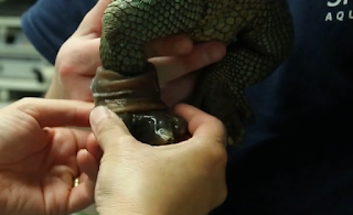
VETERINARIANS AND DESIGN TEAM GIVE LIZARD 3D PRINTED PROSTHETIC LEG.

Whenever someone asks exactly what it is 3D printing is doing that’s so important, I’m torn between which examples to share first. With impacts occurring in so many sectors, from the space industry to automotive and construction, and far more, there are countless stories offering evidence as to exactly why this technology is important. The medical realm, however, is where you find the most compelling cases, and if you are one of those individuals today who have had their lives improved or that of a baby or family member saved, then what 3D printing means to you is something most of us can’t even imagine. People around the world are reaping the benefits, as well as discovering an endless outlet for creativity. But while the human element and quality of life is a strong focus, certainly don’t discount our priority on making sure that pets and wildlife get in on this goodness too. 3D printed prosthetics in the human realm are becoming much more accessible and affordable thanks to volunteer groups like e-NABLE, providing replacement limbs for children in need all over the world from Uganda to Ghana and far beyond. And should you think that the goats, sheep, chickens, kitties, cockatoos, and dogs are being forgotten, well check earlier posts. Now, that long list has a new favorite in the form of a semi-aquatic caiman lizard who, sadly, lost a rear leg to cancer last year. lizard Hiss Majesty is 16 years old and has a devoted family at the Shedd Aquarium in Chicago. In trying to re-create a new leg and foot for this amazingly adorable creature—who patiently sits while they cover his foot in molding materials—the team, consisting of 3D designers, animal care specialists, and veterinarians, is certainly exhibiting a true labor of love, because it would seem that getting the perfect fit for those stubby little legs is not an easy task. They’ve tried ten designs so far for the right rear foot, with trial and error going strong, but with improvements in each iteration. Translating the good rear left leg into a shape for the missing one, the team has used molding techniques and experimented until they have a nice collection. The use of flexible plastics that they have created so Hiss Majesty might have better mobility seemed like a likely choice for a prosthetic, along with the use of very lightweight silicone. The most recent top pick for Hiss Majesty is a 3D printed model with perfect chunky little toes and a prosthetic insert that’s like a sock for a nice tight fit. He’s a quiet guy, Hiss, and the team isn’t sure how much the prosthetic, or the one they pick for him, will improve his life, but they are certainly going the extra mile at the 3D printer with the goal of fabricating a prosthetic that has as much joint-like movement as possible. With eventual plans to change his name to ‘Bionic Hiss Majesty,’ they continue to forge ahead and it would seem they are copiously honing their design and printing skills for the reptilian world. “The goal for this is to see if we can get something that will actually work and use that as a tool in the future to improve animal care,” said designer Kristen Neria, who helped develop the prototypes. This last prototype is much more flexible and lifelike—and promising—but they are continuing to explore the use of molds and 3D printing technology for further, even more advanced designs. Neria says their priority is definitely on emphasizing the movement of joints, and they may even start experimenting with titanium. Hiss Majesty certainly has a twinkle in his eye and while everyone is hard at work trying to give him the prosthetic of the future, it would seem he’s pretty happy with the amount of attention he’s getting in the process, whether his foot is encased in plastic, silicone, or nothing at all. [Source: Chicago Tribune]

Saturday, July 2, 2016

The new rave; 3D PRINTING.

The world is changing,many platforms for growth and development are evolving and joining the train is only sensible. The world of 3d printing is so enormous and the influence in all sectors of the economy makes it a rallying point for the business-oriented individuals. 3d printing has helped in medicine, telecommunication,food industry,clothing, jewelry,cars e.t.c the list is endless and the impact is so amazing. There are surgical procedures that could not be achieved some years back,but thanks to 3Dprinting,lives are saved. This is an open invite to catch the vibe,join the buzz,3d printing is the game-changer. The latest news from the 3d printing world according to 3Dprint.com is that the University of Sheffield is making 3D Printed Micro-Rockets Fueled for Thrust by Enzymes to Deliver Drugs in Human Body.Researchers from Chemical and Biological Engineering at the University of Sheffield are working on it and the process of delivery is astounding. University of Sheffield are responsible for having created a powerful drug delivery system via 3D inkjet printing, working on previous conventions that due to the tools at hand were just too ‘laborious’ to offer great future potential. The use of reactive inkjet printing (RIJ), the researchers expose two different solutions to each other, thus creating a new one or causing a change in form—and here the main result is autonomous, silk-based rockets with varied catalyst distribution and direction of movement. This new technology, technique, and findings, are explained thoroughly in ‘Reactive Inkjet Printing of Biocompatible Enzyme Powered Silk Micro-Rockets,’ by David A. Gregory, Yu Zhang, Patrick J. Smith, Xiubo Zhao, and Stephen J. Ebbens—just published in small. While this concept is not new and studies regarding this basic idea have been going on for at least a decade, University of Sheffield researchers have found a way to streamline it substantially with 3D inkjet printing. As interest has grown in seeing this technology progress, the focus is on applications such as:1)Environmental monitoring and remediation 2) In vivo drug delivery and repair 3)Lab on a chip diagnostics With RIJ technology, there’s much greater potential as compared to lithographic fabrication processes currently ongoing in labs. Through employing the world of 3D technology and printing, they are able to make micro-rockets, composed of silk scaffolds, and ‘highly biocompatible and non-biofouling.’ At 300 microns in length and 100 microns in diameter (as thick as one human hair), the mini-rockets autonomously propel themselves powerfully but they do require bio-fluids acting as their ‘fuel.’ This could have substantial implications in cancer treatment, as feasibly the 3D printed rockets could seek out and destroy cancer cells without causing any detriment to the human body. Not only that, more progressive benefit is found in using this new technology as it’s more affordable than trying to use devices like polystyrene beads, carbon nanotubes or metal, which require a coating such as platinum, and obviously lead to concerns regarding safety within the body. The inket printer’s ‘material’ consists of dissolved silk combined with an enzyme, presented in single droplet form by the MicroFab ‘Drop on Demand’ printer, relying on Jetlab software, and employing four single nozzle print heads (60 μm diameter) which are each attached to their own individual reservoir. The 3D inkjet printer builds up layers of ink, and they are responsible for making a column of the rocket. Beyond that, the researchers have found that the secret is then is exposing the silk to methanol which converts it into a secondary structure allowing for the capture and retention of the enzymes within. The enzyme is then what reacts and acts as the catalyst, with bubbles simply propelling the rocket. Using the silk scaffolds also eliminates the need for surfactant additives. The process is safe, biodegradable, and affordable, offering the true possibility for realistic use in applications such as drug delivery, tissue engineering, and enzyme immobilization. And as the researchers state in conclusion: “…evidenced by much recent research attention, the future potential to further develop these micro-rockets is significant.”

Sunday, June 26, 2016

Separating Conjoined Twins with the Aid of 3D Printing.

Surviving birth is already an achievement for them, but separating conjoined twins is only possible if they are not sharing vital organs. This was the situation twin girls Knatalye Hope and Adeline Faith were facing. Until they were 10 months old, they were conjoined at the chest and abdomen, which included their chest wall, lungs, pericardial sac, diaphragm, liver, intestines, colon and pelvis. Surgeons at the Texas Children’s Hospital started planning the highly-complex separation surgery nearly a year in advance. To visualize the intertwined organs, they used Materialise’s Mimics software to design a 3D-printed model based on high-quality CT scans which were designed to generate optimal contrast within the shared organs and the vasculature. Following computerized segmentation of the anatomy, the color-coded output was subsequently exported for 3D Printing and showed in great detail the babies’ heart, lungs, stomachs and kidneys, and where exactly they were connected. “Having a 3D-printed model gives you an insight into what you’re going to encounter,” Dr. Rajesh Krishnamurthy, chief of radiology research and cardiac imaging at Texas Children’s Hospital, said in a video released by the hospital. “This type of surgical planning becomes very important when you decide to assign an organ to one twin or the other.” The key collaborators in this project were Mr. Nicholas Dodd, an advanced visualization expert at Texas Children’s Hospital, and Dr. Jayanthi Parthasarathy of MedCAD in Dallas, who supervised the 3D Printing process. In addition, the surgeons implanted tissue expanders into their torsos, stretching the skin ahead of the separation. The final surgery, which took nearly 30 hours and involved more than 26 clinicians of 13 different specialties, managed to successfully separate the two babies. Contributed by materialise

Mayo Clinic Helps 11-Year-Old with Blount’s Disease by Using 3D Printing.

For 11-year-old Amarachi Austin-Okoh, running, jumping and even walking was a struggle. She suffered from a condition called Blount’s Disease, where the tibia, or shin bone, doesn’t grow properly, causing the legs to develop a bow shape. The disease had progressed so far in Amarachi’s case that even walking caused her great pain, and she explained that “It was very painful and hard, and, then, if people were walking a distance or something, I would start walking slower and slower, because it got harder and harder.” Her family had already noticed the condition when Amarachi was just two years old, but despite having a few corrective surgeries in Nigeria, where the Austin-Okoh family is originally from, her legs continued to worsen. The family approached Mayo Clinic in Minnesota to see if they could make a difference. The doctors at the Limb Lengthening and Regeneration Clinic knew they could help Amarachi if they took a team approach to the problem. By making full use of Mayo Clinic’s 3D Anatomic Modeling Lab, they were able to print out exact models of Amarachi’s leg bones to make crucial decisions prior to surgery. Dr. Todd Milbrandt, the surgeon who operated on Amarachi, was able to use the model to figure out where he would make a cut in the tibia, so that the bones could regrow and align properly. During the operation, he knew what to expect due to the pre-operative planning, and made a cut just below each knee. Dr. Andrew Sems then attached external braces (or “fixators”) to Amarachi’s legs after the operation and carefully adjusted them over the next three months according to computer calculations. By making adjustments to the fixators, Dr. Sems was able to gradually straighten the bones – basically by growing and correcting the bones at the same time. The outcome was everything Amarachi and her family could have hoped for – she gained almost 25cm in height and now walks with straight legs and no pain. She concluded, “I’m excited now, because it has opened a whole new horizon. I can do anything and everything I want to do.” Contributed by materialise

Wednesday, June 22, 2016

3D Life Makes High-Quality 3D Printed Medical Models So Doctors Can Save Lives.

3D Life is certainly aptly named. This Greek startup isn’t promoting a fun new lifestyle or arts and crafts in the 3D printing industry—they are involved in the serious business of helping to save lives. Recognizing the value that 3D printing has in the medical arena, the team at 3D Life is committed to making 3D printed models for medical professionals to use, with numerous benefits for all involved. Currently, the Athens-headquartered company makes high-quality, detailed models of the anatomy like teeth, hands, and bones, as well as organs like the heart, liver, brain, and more. 3D Life is encouraging the more comprehensive study of anatomy, mainly in terms of organs. They believe that even better knowledge of organs allows for better preparation for surgeries that are often solving complex physical issues—and the team uses congenital heart disease as a perfect example. “As a result, the doctors can better plan reparative operations based on conventional medical imaging which is suboptimal,” Bilalis told 3DPrint.com. “The human mind can only partially understand trying to create mental images of three dimensional structures so we believe that having this ability will make operations shorter and more efficient, and provide better results.” At 3D Life they believe, and undoubtedly are correct, that the exercise of holding and manipulating a 3D model leads to a better understanding of the client’s condition, as well as allowing for practicing for surgeries that are intricate and may not even have been performed before. This leads to numerous positives, from safety to better client outcomes, along with providing training for students. While there may be other companies around the world making 3D models, 3D Life is unique as the first company to endeavor in such a field in Greece. Offering advanced 3D printing, they are able to make models in a variety of materials and, even better, multiple colors. They are able to offer excellent services to all the professionals who come to them. 3D Life uses Materialise Mimics software, made specifically for medical image processions. This enables the conversion of MRIs or CTs into 3D models, which can then be used in numerous ways. These devices avail, medical professionals and surgeons to find themselves with a much better way to educate patients and their families about medical conditions, handle diagnoses and treatments, and explain procedures. Surgeons can then spend a lot of time themselves with those medical models, considering and practicing for upcoming operations—as well as using the models in the operating room to navigate through surgeries, saving time and allowing for better outcomes with less surprises. In some cases around the world, 3D libraries are being made as these models begin to pile up, and can be of use to other medical personnel. Contributed by 3D Print

Utah Woman Beats Kidney Cancer Thanks to Progressive Doctors & 3D Printing.

The advent of the 3D printed medical model, many patients and their families now are afforded a look at exactly what’s going on, thanks to completely patient-specific models made from MRI or CT data. In Linda Green’s case, not only did the 3D printed model show her the stubborn tumor lodged inside her kidney, but it may be the reason she is alive today, with the tumor that was sneakily tucked underneath her ribs now a thing of the past The 3D model is not of course just a wonderful educational tool for a patient like Green but it can serve as an invaluable guide for surgeons like Dr. Jay Bishoff. This technology and these incredibly customized visual aids previously not available to doctors, treatments can be decided on with more facts and information in front of them, literally, and procedures that have not been tried before are now possible. Not only that, doctors can train on these aids as well, a new quotient in the treatment process which doesn’t leave surgeons or medical students having to be resourceful for training devices, or spending any more time than possible practicing on cadavers. “We could not appreciate the peak of the tumor that was growing up into the drainage system of the kidney until we did the 3D reconstruction and 3D printing,” Dr. Bishoff said. For Green’s impending treatment and surgery at Intermountain Medical Center, Dr. Bishoff had a clear model made of the kidney and tumor. This allows for surgeons to have a comprehensive view and to avoid the major mistake of removing something vital during the surgery. With the intricate and transparent model, however, Dr. Bishoff was able to remove the tumor without issue, leaving all vital parts intact. He used multiple 3D prints as guides to navigate through the operation, something numerous surgeons are doing today if they have access to the extremely helpful technology. “I could’ve ended up with infection across my outer body or bleeding out,” Green said, definitely understanding the gravity of the procedure. “When the surgery was over and he came out to talk to me, I really thought he was going to tell me that he had to take the kidney out,” Green’s husband said. In a wonderful success story, Green is happy back at her home in Utah, cancer free, and her kidney is completely intact and functioning. The doctors have said that she won’t suffer any damage to the kidney or run the risk of failure. Contributed by 3D print

3D Printing Give Baby Born with Severe Cranial Defect a Future.

Bentley Yoder was never expected to survive. His mother, Sierra Yoder of Sugarcreek, Ohio, was well aware, when she went into labor on Halloween night 2015, that she would likely only get to hold her son for a few minutes before he passed away. She had known since the 22nd week of her pregnancy, when she went in for her normal ultrasound, and the doctor told her that something was very, very wrong. Something was wrong with the baby’s head, he told Sierra and her husband, Dustin, and sent them to a hospital for further tests. Neurosurgeons at the Canton hospital told the couple that their baby had a rare congenital condition called encephalocele, meaning that a portion of his brain was growing outside of his skull. About 375 babies, or one in 10,000, are born with the condition each year, according to the Centers for Disease Control and Prevention, and while there are varying degrees of severity, the disease, needless to say, causes serious difficulties for children suffering from it: developmental delays, vision problems, seizures, and more. Bentley’s case fell into the severe category, meaning that he had a very slim chance of surviving long after his birth. Even if he did live, doctors said, he wouldn’t have any cognitive function. The Yoders were encouraged to think about abortion, and they agreed to terminate the pregnancy, not wanting their child to suffer when there was no hope of recovery. The night before the scheduled procedure, however, Sierra realized she couldn’t go through with it. She and Dustin agreed on a name for the baby – Bentley Ross Yoder – and doctors gave them brochures for funeral homes in the area. Nine hours after she went into labor on Halloween night, Bentley was born. His condition was immediately clear from the massive protrusion at the back of his head, but to his parents, he was perfect 36 hours later, however, family members were still holding the baby and passing him around. The doctors, unsure of how much longer Bentley would live, told his parents to take him home and arrange hospice care, which they did. Four weeks later, they took him to a specialist at Nationwide Children’s Hospital in Columbus. The specialist took some MRIs and told the Yoders that Bentley’s brain was too damaged for him to survive much longer. Four months later, the couple took Bentley to the Cleveland Clinic. For the first time, a surgeon gave them a sliver of hope – Bentley was using his brain, which had been already clear to Sierra and Dustin. Their baby, who doctors had said was going to be born a “shell,” unlikely to ever move or even breathe, was acting just like a normal baby – indistinguishable from their first son, Beau, other than the protrusion on his head, according to Sierra. The surgeon told them it was possible that Bentley’s brain could be placed inside his skull, though she didn’t know if he could survive the surgery.. The Yoders were referred to Boston Children’s Hospital, where they met with chief plastic surgeon Dr. John Meara and neurosurgeon Dr. Mark Proctor, who had plenty of experience with cranial deformities – and with 3D printing. The two surgeons were part of a team that reshaped the skull of a baby named Violet in 2014, in a surgery that was carefully pre-planned using 3D printed models. Drs. Meara and Proctor took the same approach with Bentley’s case, 3D printing models of his cranium and planning the delicate procedure that would allow his brain to be encased within his skull. According to Dr. Meara, Bentley had 100 cubic centimeters of brain outside of his skull – a significant amount that would require his cranium to be expanded for the tissue to fit inside. Using the 3D printed models, he and Dr. Proctor devised a plan to make several vertical slices in the cranium and insert biocompatible, dissolving plates to hold it open. The protruding part of Bentley’s brain – the portion that controls vision, motor function and problem-solving, would slip inside where it belonged. yoder On May 24, 2016, Bentley went into surgery. The surgeons drained excess cerebrospinal fluid from his brain, made the cuts in his cranium, and gently eased his brain inside. Then they closed the gap using leftover bone from the cuts. Five hours later, Sierra, Dustin and Bentley’s brother Beau went to see him in the recovery room. The delicate mass at the back of his head was gone, and now, a month later, his blond curls are growing back in. He’s also holding his head up, eating, smiling and chattering like any seven-month-old. It’s uncertain what life will be like for him as he grows up, but he will grow up, and the doctors told Sierra that they believe he will have a “rewarding life,” despite any challenges or complications may arise. Using 3D printing to successfully operate on a condition as severe as Bentley’s is a new phenomenon, and therefore lacks precedence to give doctors a clear idea of what lies ahead. Bentley himself has made it clear that he is a survivor, however, and Sierra said that a part of her always knew that Bentley was going to defy expectations. That’s why she decided not to end the pregnancy. Contributed by 3Dprint

Tuesday, June 7, 2016

University of Illinois Veterinarians Use 3D Printing to Help With Eagle’s Surgery.

3D printing technology is helping veterinarians prep for surgical procedures on one of the most prized bird species in the world. Recently, students from the University of Illinois College of Veterinary Medicine had come across a wild eagle with its left humerus out of alignment, due to improper healing after it was shattered by a gunshot wound months earlier. In order to help the bird take flight again, an intensive surgery was required. To do this, the veterinarian students turned to those in the College of Engineering, who assisted by creating two 3D printed life-sized models of the eagle’s humerus, one that was healthy and another that replicated the actual injured bone. Before the the 3D printed models were used by Dr. R. Avery Bennett, an acclaimed avian surgeon, to help perform the procedure, a massive dataset was procured from the spiral CT scan taken by veterinary radiologist Dr. Stephen Joslyn. Consulting from Australia, Dr. Joslyn added a so-called ‘threshold’ into the data, which enabled the computer to separate ‘bone’ and ‘not-bone’ from the subtle and delicate CT scan information. Since the injured bone was fragmented, and thus unable to be printed in a single piece, medical illustrator Janet Sinn-Hanlon utilized software to manually thicken and link the bone areas together. After communications went back-and-forth between experts across the world, which were facilitated by Wildlife Medical Clinic intern and University of Illinois student Dr. Nichole Rosenhagen, it seemed that the life-sized models were set to be 3D printed in the university’s Rapid Prototyping Lab. But, the day before the surgery was planned, it turned out that the 3D printing queue was full. Thanks to Ralf Möller, the lab supervisor and director of technical services in the Department of Mechanical Science and Engineering, the models were 3D printed overnight in about six hours, and were good to go by the time the lab opened up the next morning. Möller enlisted the help of undergraduate student and lab technician Nick Ragano, who visited the lab overnight to ensure that 3D models would be prepared for use, and also pressure washed the starch-based support material used to print the plastic models. That morning, the 3D printed bones were collected by Dr. Rosenhagen, and the injured eagle received successful orthopedic surgery in a matter of three hours culled from 3dprint.com

Monday, May 30, 2016

Heriot-Watt University Researchers Win $3.3 Million Grant to 3D Print Smart Rocks to Capture Data on Underground Oil and CO2.

In order to secure our water, food and energy supplies while being able to maintain the safe extraction of oil and gas from underground oilfields, we need to have a better understanding of the layer of porous rocks in the subsurface. Specifically, the way that liquids and gases manage to travel through them, and how captured carbon dioxide (CO2) could be stored underground. Unfortunately the conditions of this subsurface material vary widely depending on the type of rock, the temperatures and the pressures that occur deep under the ground. Beyond the complexity of replicating specific environmental conditions, direct dynamic observations at the pore level are virtually impossible in a lab setting. According to Professor Mercedes Maroto-Valer, holder of the Robert M Buchan Chair in Sustainable Energy Engineering at Heriot-Watt University , the problem is that the rocks are unable to tell us what’s happening to them. However Maroto-Valer and her team of researchers think that they have come up with a way to communicate with these subsurface rocks, the team wants to make their own rocks that are capable of communicating with us. Their research was promising enough that the team received a prestigious European Research Council Advanced Award so they can continue to pursue their research into developing 3D printed “smart rocks” capable of giving the team an inside look of what’s happening deep underground. Maroto-Valer and her team will use a 3D printing process to produce their own porous rocks that will include multiple micro sensors embedded inside of them. The sensors will be able to transmit data directly to the research team, including detailed information about what actually happens to liquids and gases deep underground. The 3D printed smart rocks will be capable of providing information on the subsurface conditions and environment at a microscopic level, which is simply not possible to reproduce using traditional laboratory methods. This fundamental knowledge at such a tiny scale will feed hugely into our understanding of such processes at the large scale and enable us to maximize the success of industries from oil extraction to water safety and the storage of captured CO2. The grant that Maroto-Valer and her team won was awarded by the “Excellent Science” pillar of Horizon 2020, the European Union’s research and innovation program focusing on enabling senior researchers to pursue their most promising ideas. read more at 3dprint.com

Friday, May 20, 2016

3D PRINT RECYCLING.

One of the downsides of 3D printing is trash. This does in fact occur mainly by way of plastic—and even just one home workshop can produce surprising volumes of it, by way of big blobby print failures, discarded prototypes, and other materials as well. Upon the advent of this technology, most were caught up in the euphoria of discovering something new that allows for such empowerment in making things, verging on that of magic. It wasn’t too long, however, before the question of what to do with piles of plastic in the trash emerged. Lately, we discover and discuss numerous ways of recycling, as well as using recycled products in filament that can then be recycled again—and even for solvents. Companies like Voodoo Manufacturing and Filabot are concerned about trash pileup too, and rather than just setting that worry aside, they have taken action in creating a partnership for recycling. This is where the fun comes in—well, maybe it’s just me—but the idea of chucking all that plastic into a great big grinder and sending it back full circle seems very fulfilling. Plus, let’s consider the savings on the bottom line here. Filament, while varying in price, can begin to take a toll on the budget if you are a major 3D printing enthusiast. Voodoo and Filabot have discovered quite a symbiotic relationship. Voodoo has bunches of plastic that needs to go out the door and Filabot has the means to recycle and re-use. This is a real rags to riches story for trashed items that are turned back into what is often quite an expensive commodity. The Filabot machinery can recycle practically any plastic, grinding it, and sending it right back where it came from, also coming full circle in some of the most wonderful 3D printing benefits—self-sustainability, innovation, and affordability. Have you been worried about what to do with plastic from 3D printing? recycle is the answer. read more at 3Dprint.com

Sunday, May 8, 2016

The Polysher will shine your 3D-printed objects.

Polymaker wants to make 3D printing a tempting hobby for everyone with its new PolySmooth filament and Polysher machine, which polishes creations so they look shiny and cohesive. The company launched the items on Kickstarter this past week and has already surpassed its $100,000 goal. The filament can go in any extrusion-based 3D printer, but the Polysher will only work on items that have been made with PolySmooth. The polisher works by simply spraying the piece with an alcohol aerosol solution. Polymaker hopes nicer looking 3D objects will bring 3D printing into mainstream culture. No one currently mixes up mass-produced plastic pieces with printed ones because of their feel and obvious filament layers. Solely going off Polymaker's Kickstarter images, the new filament and Polysher really does make 3D printer creations look more legit. There are no obvious layers, and they come off as fully finished products. I'm not sure if the Polysher will finally bring 3D printing into the mainstream, but in either case, I'm always pro-shiny things. culled from polymaker.

Sunday, May 1, 2016

3D-printing dentists are recreating entire jaws.

3D-printing has been used for basic dental procedures for years – and the technology is now advanced enough to reconstruct entire jaws. Andrew Dawood, a dentist with Dawood and Tanner, told the audience at WIRED Health that they have used the lost wax technique which has been around 5,000 years,but 3D-printing has allowed him to do more with patients and do it faster. Dawood explained that he was able to design and print the frameowrk, cutting guides and parts needed to perform complex dental surgery. He said all it required was data collected from 3D scans and patient records. Plaster of Paris parts used for surgery are now 3D-printed and Dawood also uses plastics, rubbers, and metals while printing. He said they are not 3D-printing bone yet,but will one day. A technique to make and 3D-printed human bone hasn't been developed yet, but Dawood and other surgeons are already using bone from other parts of the body to reconstruct entire jaws in the most complex cases. Josh Stephenson, a designer and one of Dawood's patients, had his jaw reconstructed after suffering from a malignant melanoma. After unsuccessful radiotherapy treatment, Stephenson underwent surgery to remove his left eye, upper left jaw and the roof of his mouth. Using a 3D-scanned and printed copy of Stephenson's skull it was possible to recreate the missing parts of his jaw. Stephenson, who is a graphic designer, has since used the same 3D-scanning and printing technique to create new products. Dawood was able to asses the dentistry work needed from the 3D-printed model and then say how surgery to repair it should be carried out. Using "bone taken from [Stephenson's] scapular" the dentist and other surgeons reconstructed his jaw. Using this type of technology, there can be more accurate intervention and quick return of patient to society . culled from wired.co.uk

Tuesday, April 19, 2016

3D PRINTING AND VETERINARY MEDICINE.

In PennDesign’s Fabrication Lab, students and faculty use three-dimensional printers to craft geometric forms, architectural models, and other products of the imagination. But in a recent collaboration with the School of Veterinary Medicine, the printers have been put to work making models based very much on reality. After examining a skull deformity afflicting a canine patient named Millie, Evelyn Galban, a neurosurgeon and lecturer in Penn Vet’s Department of Clinical Studies-Philadelphia, thought it would be useful to physically handle a replica of the dog’s skull. “It’s difficult to fully understand the malformation until we have it in our hands,” she says. “That usually doesn’t happen until we’re in surgery.” The expertise of PennDesign’s Stephen Smeltzer and Dennis Pierattini partnered with Galban, along with veterinary neurology residents Jon Wood and Leontine Benedicenti, to produce models that precisely replicate injuries or deformities of pet dogs and cats. These applications have the potential to improve training and patient care at Penn Vet, while stretching the imaginations of PennDesign students and faculty. Pierattini remarked they are very interested in finding more ways that can explore the potential of the equipment and fathom its depths. The veterinarians took CAT scan, then transformed CAT scan files into a format that the 3D printers could recognize.They produced the skull of Millie, composed of gypsum powder bound by acrylic and sealed with a super glue-like substance to make it rigid. These models could help vets like Evelyn Galban plot out and practice surgical procedures in advance of an operation. Full-color models may even allow for testing new approaches that avoid contact with critical blood vessels and other tissues. Read more;
http://www.upenn.edu/spotlights/3d-printing-veterinary-surgeries

Agribusiness ideas.

Agribusiness Millionaires

Agribusiness Millionaires
Learn how to make money in agribusiness.

Popular Posts

AGRIBUSINESS EDUCATION.

Translate

I-CONNECT -AGRICULTURE

AGRIBUSINESS TIPS.

AGRIBUSINESS.

The Agriculture Daily

veterinarymedicineechbeebolanle-ojuri.blogspot.com Cassava: benefits of garri as a fermented food. Cassava processing involves fermentation which is a plus for gut health. The fermentation process removes the cyanogenic glucosides present in the fres...

Claim your bonus here..

Claim your bonus here..
Free dog care guide.

CASSAVA BUSINESS

CASSAVA BUSINESS
CASSAVA FLAKES.